
Ultrasound scanning is an important clinical tool in providing images of internal fetal anatomy. It is also called sonography because it uses high-frequency sound waves to produce images of slices through the body. A transducer or probe which emits ultrasound waves is placed on the skin after coating it with a thin layer of conductive gel, to make sure the waves pass smoothly through the skin. The emitted ultrasound waves are reflected by different structures encountered by the waves, the strength of the reflected waves, and the time they take to return form the basis for interpreting the information into a visible image. This is performed by computer software. The advantages of ultrasound imaging over other imaging techniques include:
 Real-time visualization of the fetus or organs.
Non-invasive.Doesn’t use ionizing radiation, which has been associated with toxic effects on the embryo.Doesn’t use ionizing radiation, which has been associated with toxic effects on the embryo.
Real-time visualization of the fetus or organs.
Non-invasive.Doesn’t use ionizing radiation, which has been associated with toxic effects on the embryo.Doesn’t use ionizing radiation, which has been associated with toxic effects on the embryo.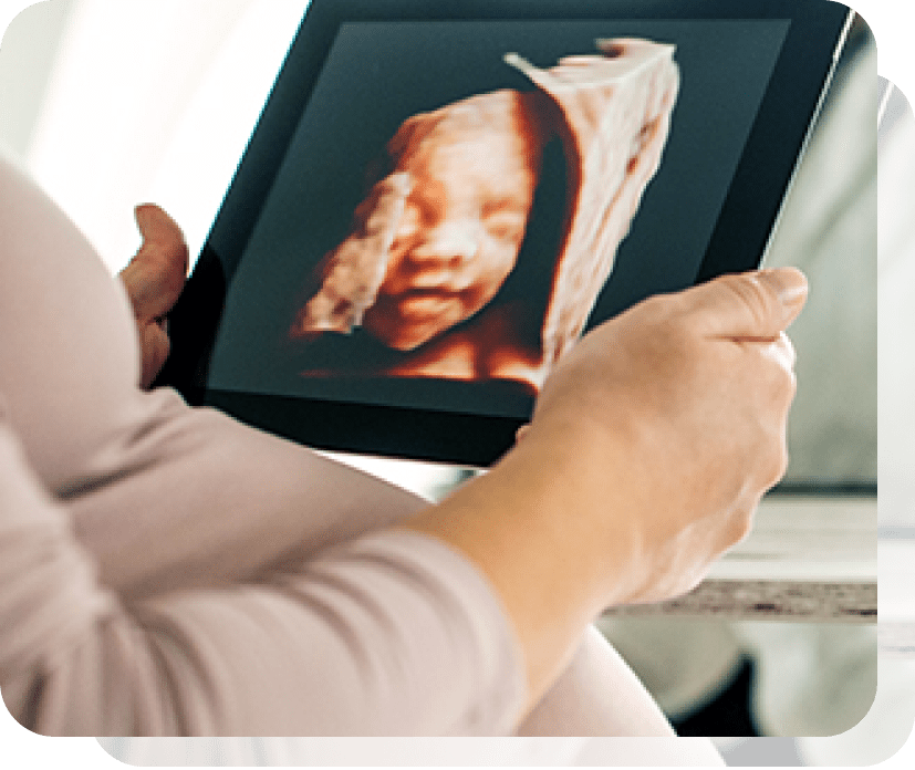
2D Ultrasound sends and receives ultrasound waves in just one plane. The reflected waves then provide a flat, black-and-white image of the fetus through that plane. Moving the transducer enables numerous planes of viewing, and when the right plane is achieved, as judged by the image on the monitor, a still film can be developed from the recording.
Further development of ultrasound technology led to the acquisition of volume data, i.e., slightly differing 2D images caused by reflected waves that are at slightly different angles to each other. These are then integrated by high-speed computing software. This provides a 3-dimensional image.
The use of virtual planes helps in better visualization of fetal heart structures by allowing views not attainable by 2D imaging, possibly adding a 6% chance of detecting defects.Diagnosis of fetal face defects like cleft palate.Diagnosis of fetal skeletal or neural tube defects.Less time for standard plane visualization.Less dependence on operator’s skill and experience for diagnosis of common fetal anomalies.3D imaging allows fetal structures and internal anatomy to be visualized as static 3D images. However, 4D ultrasound allows us to add a live streaming video of the images, showing the motion of the fetal heart wall or valves, or blood flow in various vessels. It is thus 3D ultrasound in live motion.
Shorter time for fetal heart screening and diagnosisVolume data storage for screening, expert review, remote diagnosis as in remote areas, and teachingEnhanced parental bonding with the baby.Healthier behaviour during pregnancy as a result of seeing the baby in real-time and in 3D.More support by the father after visualizing the baby’s form and movement.Possibly more accurate identification of fetal anomalies especially of the face, heart, limbs, neural tube and skeleton.Assessment of fetal growthEvaluation of fetal well-beingPlacental localization and assessmentSeeing and hearing the fetal heartbeatCapturing images of the baby which bond the family and friends with the baby before birth